


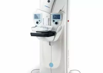
- Xenox C200 avaialble in diffrent models: with rotating anode tube or stationary one & Image Intensifier 9” or 12” (only for rotating version)
- Horizontal run 210 mm
- Arc orbital Movement 135°
- WIG-WAG ±12,5°
- C-arm depth 690 mm
- S. I. D. 988 mm/980 mm/928 mm
- Arm rotation around horizontal axis ±270°
- Generator power in DC current 5 kW (3,5kW@115 Vac)
- Generator operating frequency 40 kHz
- KV range 40 -h 120 kV
- Max. current in continuous fluoroscopy 8,0 mA
- Max. current in «SNAPSHOT» fluoroscopy 30 mA
- Max. current in HCF with DFG (HRP)30 mA
- Max. current in pulsed fluorography with DFG (HRP)60 mA@230 Vac;45 mA@115 Vac
- Max. current in digital graphy-mode with DFG (HRP)60 mA@230 Vac;45 mA@115 Vac
- Max. current in radiography (hi-rad) 35 mA @ 115 Vac ; 50 mA @ 230 Vac
- Max. mas in radiography 90 mAs @ 115 Vac -;125 mAs @ 230 Vac
- Max. Fluoroscopy Time H.U. Safety after 28 min of fluoroscopy @120 kV, 5 mA (600W)
- Membrane keyboard with alphanumeric touch-screen 5.7” LCD display for all the operative parameters and error messages. Microprocessor management. Keyboard can be rotated of ± 60°
Operating Modes and Functionality OPERATING MODALITIES OF MEMORY
- CONTINUOUS FLUOROSCOPY
- PULSED FLUOROSCOPY (2/sec, 1/sec, 1/3sec – without acquisition on hard disk; 1,3,6,12,25 fps with acquisition on hard disk)
- DIGITAL SNAPSHOT
- FLUOROSCOPY mA (1/2): (range: 0,25-4 mA)
- RADIOGRAPHY: 2 points technique (kV and mAs)
- CINE sequence: up to 25 fps. (included in basic configuration)
- Number of images on Hard Disk: about 110.000 (Hard Disk 250 GB)
- Video signal: Digital camera 1 kx1 k
- Video output: 2 x DVI 1280×1024
- Image format of the working memory: 1024 x 1024 x 12 bit
- Image format: 1024 x 1024 x 12 bit
- Number of monitors: 2-19“ LCD
- Optional Hard disks: 500 GB (About 220.000 images) or 1 TB (About 440.000 images)
SOFTWARE FUNCTIONS: selection of anatomic programs; recursive filter (1,2,4,8,16); edge enhancement Smooth, Normal, Sharp in post processing; smart filter with «motion detection»; brightness and contrast; virtual collimator, horizontal and vertical flip; electronic rotation at 1 ° step; Electronic zoom factor from 1,2 to 3; Electronic lens factor from 1,2 to 3; Overview (4,9,16 images); Text editing; Dose report; Patients archive; Interface for network Ethernet TCP/IP; Export single BMP image on USB; Cineloop rewiew; Programmed acquirement sequences: 1,3,6,12,25 fps; Grey scale inversion; max. opacity fluoroscopy acquirement; MEASURE TOOLS(included in basic configuration): length, angles, stenosis, length calibration on reference object measure; text overlay DSA TOOLS (Option): Subtraction in real time with manual/automatic mask; shifting pixels; Land Marking;
Features
- 12″ and 9″ image intensifier option available
- High Frequency monoblock X-ray generator 5 KW
- Digital Subtraction Angiography (DSA) 25frames/ses
- 1k CCD camera delivers sharp and detail-rich images
- HD memories with 350.000 images DVD, USB
- the worksation equipped with 2 LCD monitors 19″
- Modular configurations even after sales
- Full Dicom(optional)
- 200 mm horizontal C-arm run, with manual brake for locking
- Orbital movement 125°
- 270° on each side arm rotation, with manual brake for locking.
- 12° on each side C-arm swivelling with manual brake for locking.
Applications
- DSA
- Traumatology
- Orthopedics
- Digestive system
- Generic Surgery
- Biliary drainage and stenting
- Image guided biopsy
- Neonatology and pediatrics
- Lithotripsy
Options
- DICOM OPTIONS AVAILABLE: DICOM VERIFY (SCU/SCP), DICOM STORAGE, DICOM WORKLIST (SCU), DICOM PRINT (SCU), DICOM CDR/DVD, DICOM QUERY/RETRIEVE (SCU), DICOM MPPS (CPU), DICOM STORAGE COMMITMENT (SCU), DICOM DOSE STRUCTRED REPORT
- DSA TOOLS
- HARD DISK SSD 500 GB (about 220.000 images)
- HARD DISK SSD 1 TB (about 440.000 images)
- Thermal printer
- Patient radiation dose measuring device (DAP chamber)
- Laser localizer for centering the anatomical area to be examined on the I.I. side
- 24×30 cm Cassette Holder (9” I.I.)
- 35×35 cm Cassette Holder (12” 1.1.)


المراجعات
لا توجد مراجعات بعد.